Microscopy
Nikon Eclipse E-600 Research Light Microscope (LM)
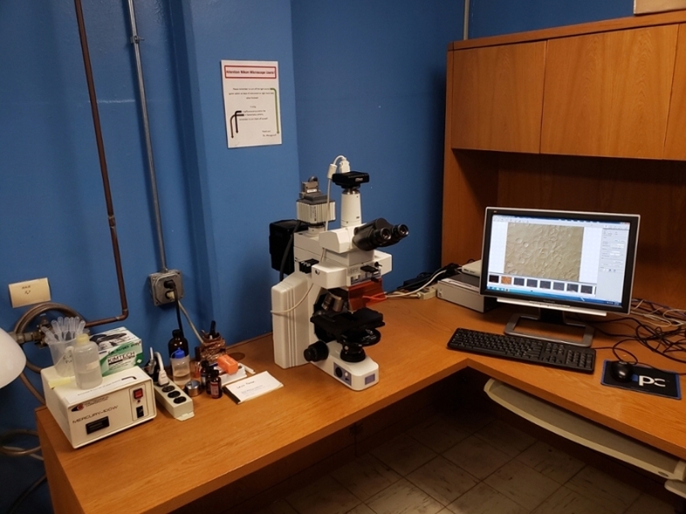
Our Nikon Eclipse E-600 LM is outfitted with standard brightfield visualization, epifluorescence filter sets (UV-2B, UV-2E/C, BFP, GFP, YFP, TRITC HYQ), differential interference contrast (DIC), phase contrast, multiple Plan Fluor and Plan Apo objectives ranging from 1X to 100X (oil), and two imaging systems: Nikon DS-Fi1c color digital camera with NIS-Elements software and Hamamatsu ORCA-ER monochrome CCD camera with HCImage Live software.
Leica MZ7.5 Stereo Microscope
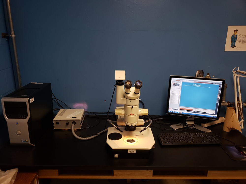
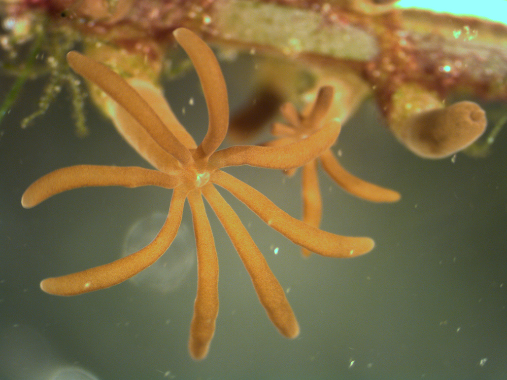
Our Leica MZ7.5 stereo microscope is outfitted with a Leica MC170 HD color digital camera, Leica Application Suite image capture and optimization software and a LAS MultiTime time lapse capture mode that can generate video files (.avi).
Zeiss LSM 5 Pascal Confocal Laser Scanning Microscope (LSM)
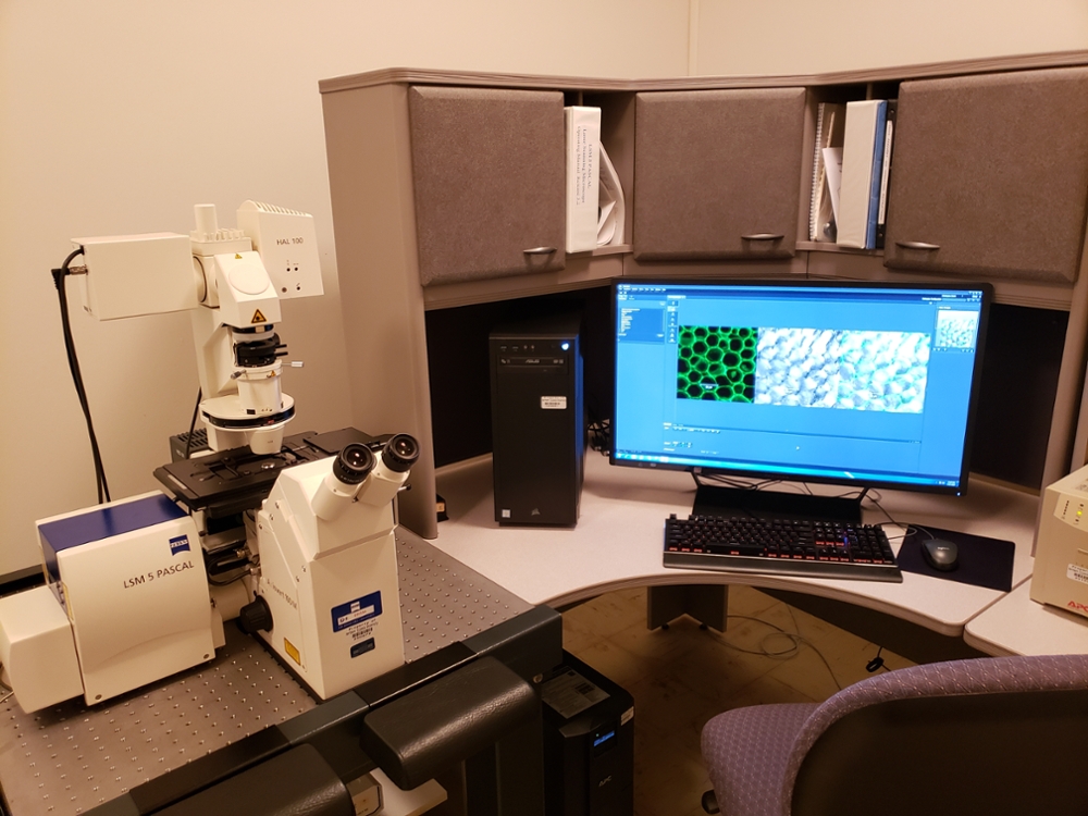
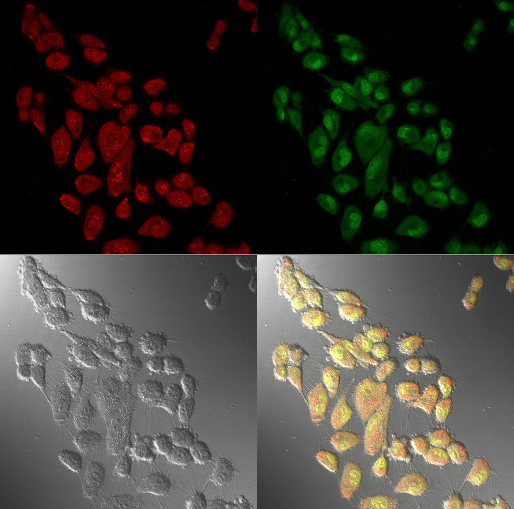
This older confocal microscope is equipped with 10X, 40X (oil), and 63X (oil) Plan Neofluar objectives, argon (458, 488, 514 nm) and HeNe (543 nm) lasers, and Zeiss LSM 5 Pascal image acquisition software.
Zeiss LSM 900 Confocal Laser Scanning Microscope with Airyscan 2
Please note: Linda Yasui is solely responsible for the management of the Zeiss LSM 900 Confocal Laser Scanning Microscope with Airyscan 2. Please contact lyasui@niu.edu for questions regarding operations, specifications or scheduling.
Additional Microscopy Resources
- Nikon MicroscopyU website
- Transmission electron microscopy equipment:
- Reichert-Jung SuperNova Ultramicrotome
- Reichert OmU2 Ultramicrotome
- Sorvall Porter-Blum MT2-B Ultramicrotomes (2)
- LKB Glass Knifemaker
- Light microscopy support equipment:
- AO T/P 8000 Automated Tissue Processor with Tissue-Tek II Paraffin Dispenser
- AO Spencer 820 Microtomes (3)
- Lab-Line Slide Warmer
- Lab-Line and Tissue-Tek tissue flotation baths
- AO Knife Sharpener
Facility Details
Director
Barrie Bode, Ph.D.Core Lab Director
Professor, Biological Sciences
bodebp@niu.edu
Location
BIOS Research Instrumentation Core (BRIC) and Core Microscopy Facility (CMF)
Montgomery Hall
Rooms 112, 113, 115, 116 and 119
Contact Us
Department of Biological SciencesMontgomery Hall 349
815-753-1753
815-753-0461 (fax)
General questions can be directed to kmeyer5@niu.edu.
Student enrollment questions can be directed to sfarley@niu.edu.