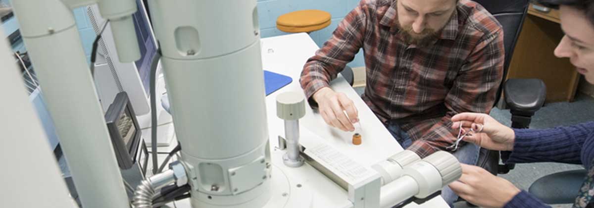
Scanning Electron Microscope, JEOL JSM-5610LV with Thermo Scientific UltraDry EDS
Description
The department's SEM has several imaging and operation modes. Realistic zoom range is from 50x to 50,000x or mm scale to µm scale objects. This varies with the type of sample and mode of operation. With the exception of "Low Vacuum Mode" below, samples need to be electrically conductive or coated in a conductive coating. The department has both a carbon coater and sputter coater. All imaging modes take place in a high or low vacuum which could damage some delicate samples.
Secondary Electron Imaging (SEI)
Used for taking high resolution images of surface details.
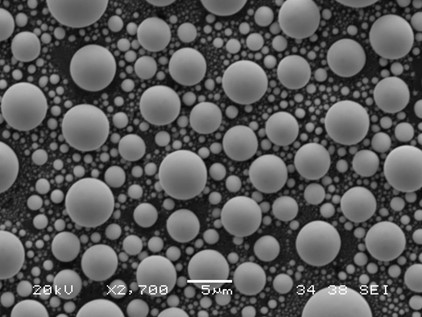
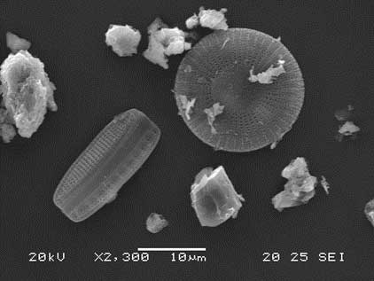
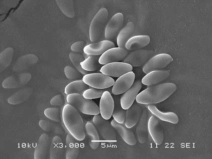
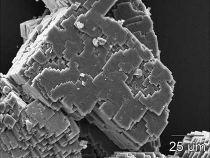
(Other examples of SEI images)
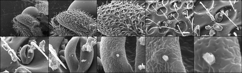
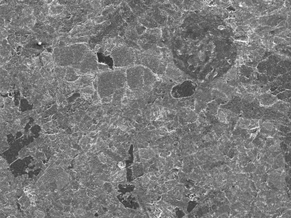
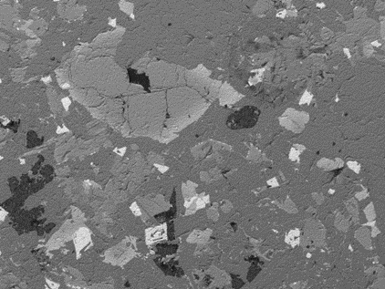
Backscatter Electron Imaging (BEI)
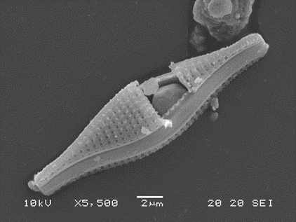
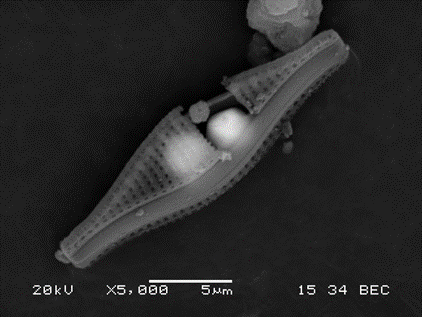
Images are created from electrons that are elastically scattered from atomic nuclei comprising a sample. In general, the denser or closer the material, the more electrons that are backscattered. This can be used to show surface detail, but it is also used to show composition changes on flat surfaces or materials just beneath the sample surface. Below is an image of a polished rock imaged in SEI (top left) and then in BEI (top right). You can clearly see the different minerals present in the backscatter image. The second set of images show a diatom with a grain stuck inside it. In SEI (bottom left) you can resolve the grain, but in BEI (bottom right) you can see through some of the top layer and see that there is also a second grain inside the diatom.
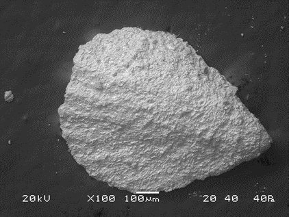
Low Vacuum Mode
This mode uses the backscatter electron detector, but allows a variable pressure in the sample chamber, which in turn, allows uncoated, non-conductive samples to be imaged. The image quality will be less than that obtained with normal SEI or BEI. Below is an uncoated brachiopod fossil.
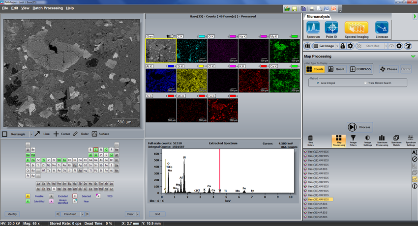
Energy Dispersive Spectroscopy (EDS)
EDS, also known as Energy Dispersive X-ray Analysis (EDX) is a qualitative or semi-quantitative elemental analysis method. The flatter the surface being analyzed, the better the results. The system can obtain the elemental spectrum from the entire field of view, spots, selected areas, lines, or create maps of elements. It can also create phase maps of similar spectra. Below is the same rock sample from above, mapped for elements.
Contact and Pricing
SEM use is charged per the hour. The cost includes normal sample preparation, which is not part of the operation time. Academics and students that wish to be trained and run their own samples may be charged a reduced rate.
| Description | NIU | External Academic | Commercial |
|---|---|---|---|
|
SEM use |
$50 |
$60 |
$100 |
|
Technician time** |
N/A |
N/A |
$100 |
Contact Josh Schwartz at joshua.schwartz@niu.edu if you have any questions.
Contact Us
Micro-Compositional Analysis LaboratoryJoshua Schwartz
Laboratory Manager
joshua.schwartz@niu.edu
815-753-7930