Image Reconstruction Software Using Proton Computed Tomography (pCT)
Principal Investigators: George Coutrakon, Ph.D., Department of Physics; and Nicholas Karonis, Ph.D., Department of Computer Science.
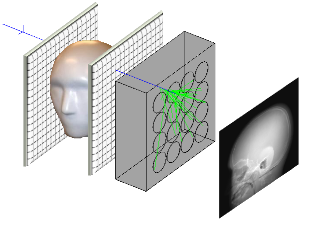
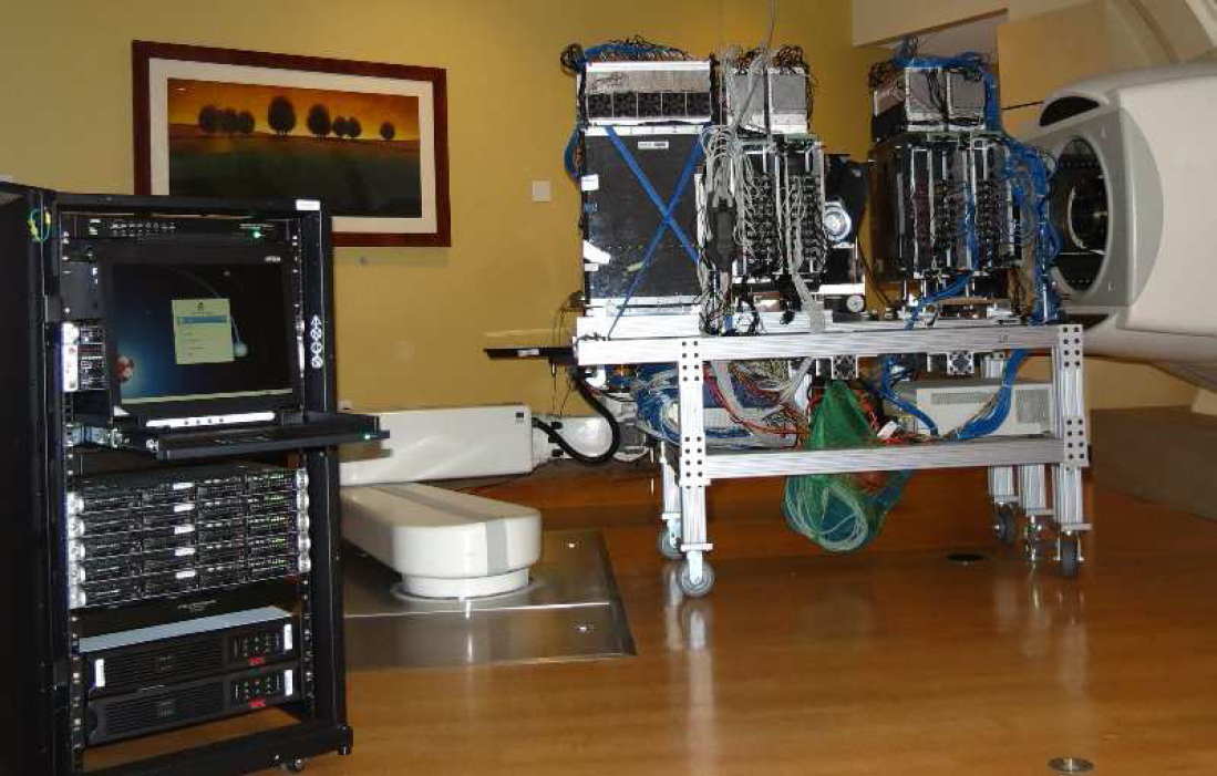
Proton imaging (i.e., radiography and tomography) is the most direct method of image guidance for proton therapy. We aim to develop a high-performance, low-cost proton imaging system based on well-established fast scintillator technology. We also aim to deliver a lower dose (relative to equivalent x-ray images) to the patient with proton imaging. This dose advantage relies on a precise reconstruction of the residual ranges of individual protons traversing the patient, and a strategy to maintain low residual range during the scan. The basic principle of proton imaging is that tracking detectors measure the position of protons before entering and after exiting the patient and that a residual range detector measures the proton energy absorbed within the patient. When imaging protons are directed through the patient in one direction we can form a radiograph image showing the proton range across the field. When the imaging protons are directed through the patient from 360 degrees around the patient we can form 3D tomographic images.
Prospective user?