Medical Physics
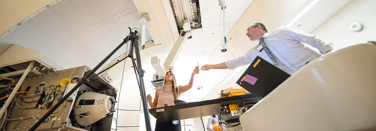
NIU is working with proton therapy facilities in Illinois and California to develop a next generation device for use in proton radiology (2D images) and proton tomography (3D images). The information provided by these imaging methods complements normal CT scans (which use photons) and MRIs.
Proton Radiology and Computed Tomography
Proton radiography can be used to verify a proton therapy treatment plan. For example, prior to a patient undergoing proton radiation therapy, proton radiography can be used to verify the treatment plan worked out earlier by a CAT scan. Proton computed tomography (pCT) can be substituted for an X-ray CT scan. It promises to provide more accurate imaging and to reduce the amount of radiation a patient receives during imaging.
NIU has helped develop two prototype devices. The first used silicon strips and was limited by proton rate. The second is a new pCT detector with the following characteristics:
- Constructed using scintillating fiber and a scintillator-based calorimeter. Both are read out by silicon photodetectors and collect data.
- Developed in collaboration with Fermilab and Delhi University.
- Supported by a grant from the Department of Defense. This grant has also funded a large GPU/CPU cluster located in Swen Parson Hall to "unpack" the data the detector collects using the proton beam at the therapy center in DuPage County.
For more information, contact faculty member George Coutrakon at gcoutrakon@niu.edu.
Contact Us
Northern Illinois Center for Accelerator and Detector DevelopmentLaTourette Hall, room 202
815-753-2700
information@nicadd.niu.edu