Irina Nesterova
Bioanalytical chemistry
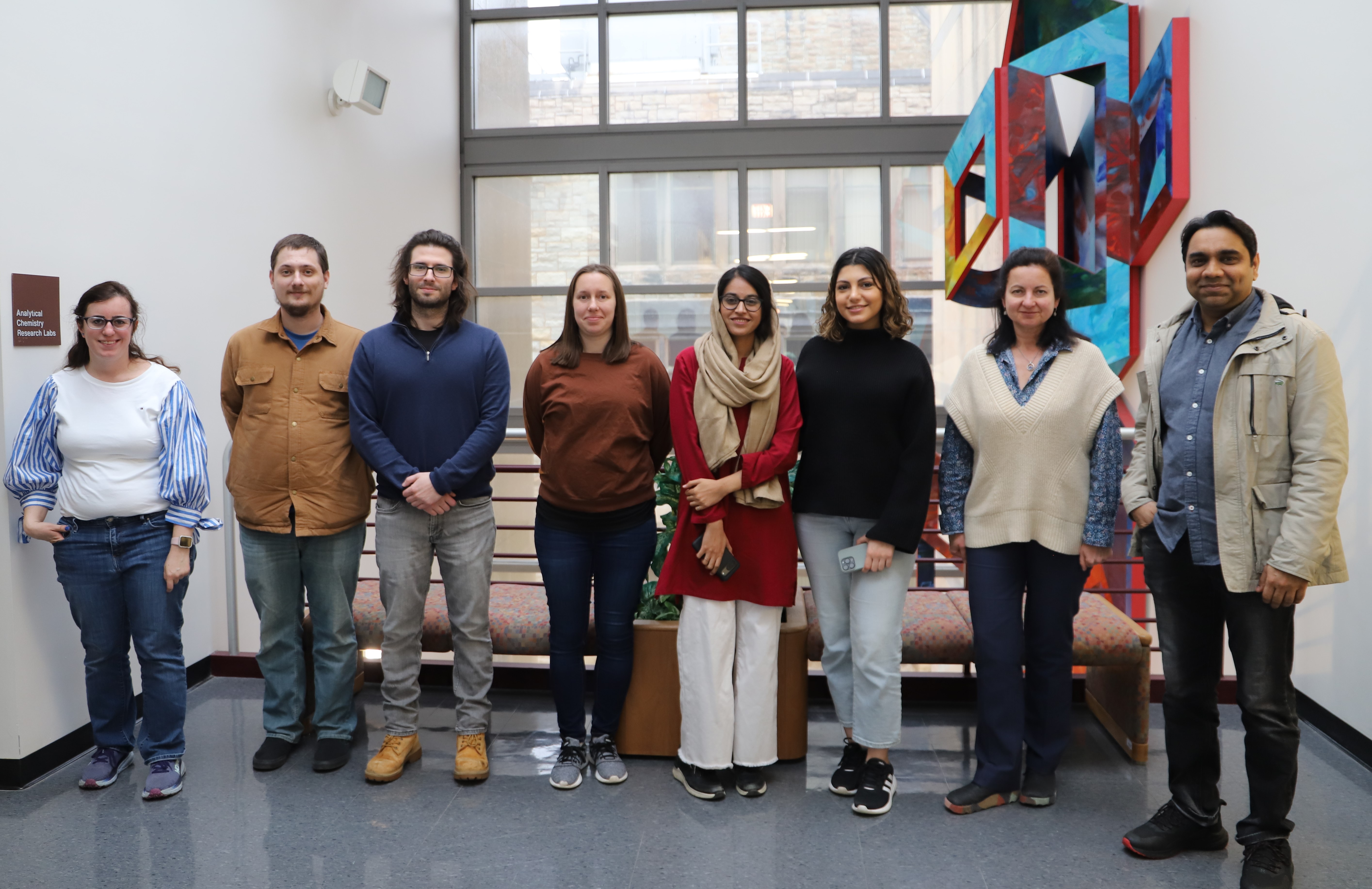
I joined the NIU Department of Chemistry and Biochemistry in Fall 2017. Currently, our group consists of several graduate and undergraduate researchers. We develop new approaches to molecular diagnostics and the analysis of living systems.
Students working in my lab will become proficient in various contemporary analytical techniques and develop a solid vision and understanding of the modern field of biological analysis. My ultimate goal is to train the students to become successful in their research field. Therefore, upon graduation, they will be well-equipped to pursue their professional dreams and compete in the modern analytical chemistry job market.
You will find brief descriptions of our research directions below but keep in mind our projects are quite dynamic and evolve much faster than we can update the page. The best way to learn about our current research is to contact me or other group members.
Research Lab

Bioanalytical chemistry: new approaches molecular analysis
Nucleic acids as functional materials
Everyone knows the primary biological role of deoxyribonucleic acid (DNA) as a genetic information carrier. Through evolution, Nature refined DNA into very stable, programmable, compatible with biological systems, and non-toxic molecules. No wonder, it is very attractive for chemists to look at DNA beyond its pure biological role and to design practically useful molecular devices.
The most common form of DNA in living system is a double helix with two complementary strands held together via hydrogen bonding between G – C and A – T base pairs. However, DNA bases can interact through hydrogen bonding which is different from classic base pairing (so-called non-canonical base pairing). Both, classic and non-canonical base pairing can lead to different folding conformations of DNA molecules (some examples are included in Figure 2). We design DNA molecules that change conformations in response to a biologically relevant signature (e.g. a presence or a quantitative threshold of a biomarker, change in heat, change in pH). Then, we convert the conformation change into an analytical signal to make molecules operate as sensors, i.e. show an easy-to-read response for the target presence or amount.
Molecular calorimeters. We are developing molecular sensors that quantify very small changes in small dissipating inputs (e.g. heat released in open systems, protons released in the buffered environment, etc.). The sensors are based on noncanonical DNA i-motifs, a single-stranded DNA that folds/unfolds in response to changes in heat or proton concentrations. To be able to detect very small signals, we make the sensors work irreversibly: they unfold in response to a change but do not fold back when the change dissipates. As a result, such sensing systems are capable of integrating small dissipating signals over long observation times as opposed to conventional sensors that follow changes in a target and return to their original state when the change disappears. Recently, we have developed the first molecular calorimeter that operates based on this principle. It measures heat released in very small volumes with much smaller amounts of binding partners compared to conventional calorimetric techniques.
Bubbling as a signal readout. The goal of this project is to develop a reliable way to visualize a response to molecular recognition events in the form of bubbles. Observing bubbles is an exciting, straightforward, and easy-to-interpret route towards instrument-free visualization of a readout that does not require scientific training, equipped lab, or color vision proficiency and, therefore, can easily extend towards mass applications suitable for everyone ages 2 and up.
To enable the readout, we develop DNA scaffolds that demonstrate catalase activity only when folded into a certain conformation. Catalases catalyze hydrogen peroxide decomposition (2 H2O2 ® 2 H2O + O2 (g)). The reaction produces O2 gas that forms bubbles. Further, we engineer a mechanism that induces the required catalytic activity conformation and, as a result, activates bubble production only when a target is present. Also, we are working on incorporating bubbling readout into quantitative analysis.
Phthalocyanine-based near-IR sensors
In collaboration with Prof. Nesterov’s group (Organic Chemistry), we develop simple near-infrared (NIR) fluorescent sensors with a specific focus on cellular applications. Our sensors become fluorescent upon binding to a protein target while remaining completely dark in the absence of the target. The turn-on response is based on the aggregation/de-aggregation mechanism of phthalocyanine (Pc) fluorophores: the Pc fluorophore stays completely non-fluorescent when aggregated in aqueous media but becomes brightly NIR fluorescent when the probe binding to a target causes de-aggregation. To enable selectivity of target binding, each sensor has an “anchor” domain that is based on a known inhibitor for the target. Our ultimate goal is to develop assays that assess small molecule inhibitors in living cells. Such assays will find applications in drug discovery as well as in the therapeutic field.
Recent publications
Minasyan, A.S.; Peacey, M.; Allen, T. K.; Nesterova, I.V. Sequence Context in DNA i-Motifs Can Nurture Very Stable and Persistent Kinetic Traps. ChemBioChem, 2024, e202400647
Mohamed, M.; Klenke, A.; Anokhin, M.; Amadou, H.; Bothwell, P.; Conroy, B.; Nesterov, E.; Nesterova, I. Zero-Background Small-Molecule Sensors for Near-IR Fluorescent Imaging of Biomacromolecular Targets in Cells. ACS Sens. 2023, 8 (3), 1109-1118
Iwaniuk, E. E.; Adebayo, T.; Coleman, S.; Villaros, C. G.; Nesterova, I. V. Activatable G-Quadruplex Based Catalases for Signal Transduction in Biosensing. Nucleic Acids Res. 2023, 51, 1600-1607
Adegbenro, A.; Coleman, S.; Nesterova, I. V. Stoichiometric Approach to Quantitative Analysis of Biomolecules: The Case of Nucleic Acids. Anal. Bioanal. Chem. 2022, 414, 1587-94
Minasyan, A. S.; Chakravarthy, S.; Vardelly, S.; Joseph, M.; Nesterov, E. E.; Nesterova, I. V. Rational Design of Guiding Elements to Control Folding Topology in i-Motifs with Multiple Quadruplexes. Nanoscale 2021, 13, 8875-83
Nwokolo, O.A.; Kidd, B.; Allen, T.; Minasyan, A.S.; Vardelly, S.; Johnson, K.D.; and Nesterova, I.V. Rational Design of Memory-Based Sensors: The Case of Molecular Calorimeters. Angew. Chem., Int. Ed., 2021, 60, 1610-4
Ducharme, G. T.; LaCasse, Z.; Sheth, T.; Nesterova, I. V.; Nesterov, E. E. Design of Turn-On Near-Infrared Fluorescent Probes for Highly Sensitive and Selective Monitoring of Biopolymers. Angew. Chem., Int. Ed. 2020, 59(22), 8440-4
LaCasse, Z.; Briscoe, J. R.; Nesterov, E. E.; Nesterova, I. V. Multidimensional Tunability of Nucleic Acids Enables Sensing over Unknown Backgrounds. Anal.Chem. 2019, 91, 14275-80
Debnath, M., Farace, J.M, Johnson, K.D., Nesterova, I.V. Quantitation without Calibration: Response Profile as an Indicator of Target Amount. Anal. Chem. 2018; 90(13), 7800–3
Nesterova, I. V.; Briscoe, J. R.; Nesterov, E. E. Rational Control of Folding Cooperativity in DNA Quadruplexes. J. Am. Chem. Soc. 2015; 137(35), 11234-7
Nesterova, I. V.; Elsiddieg, S. O.; Nesterov, E. E. A Dual Input DNA-based Molecular Switch. Mol. BioSyst. 2014; 10(11), 2810-4
Nesterova, I. V.; Nesterov, E. E. Rational Design of Highly Responsive pH Sensors Based on DNA i-Motif. J. Am. Chem. Soc. 2014; 136(25), 8843-6
Nesterova, I. V.; Elsiddieg, S. O.; Nesterov, E. E. Design and Evaluation of an i-Motif-Based Allosteric Control Mechanism in DNA-Hairpin Molecular Devices. J. Phys. Chem. B. 2013; 117(35), 10115-21

Irina Nesterova, Ph.D.
Associate Professor
La Tourette Hall 433
815‐753‐6843
inesterova@niu.edu
Research Interests
Development of sensing systems for biological analysis; methodology of bioanalysis; biocompatible signal transduction platforms; monitoring of biochemical events inside cells; and in vitro.
Educational Background
Postdoc, Louisiana State University, 2006–2012
Ph.D. (Chemistry), Moscow State University, 1997
B.S., M.S. (Chemistry), Moscow State University, 1992